Intraoperative EtCO₂ Elevation and Capnographic Abnormalities Induced by Reduced Respiratory Rate in a Dog Under Mechanical Ventilation
Abstract
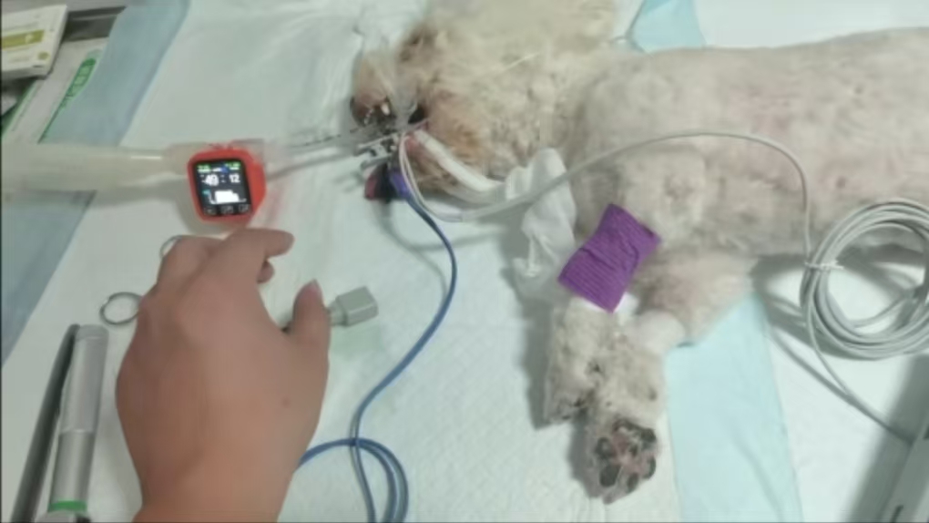
During general anesthesia in a medium-sized dog, a marked elevation in end-tidal carbon dioxide (EtCO₂) from 35–38 mmHg to 50 mmHg was observed following a reduction in respiratory rate from a spontaneous 40 breaths per minute (bpm) to a controlled rate of 15 bpm under mechanical ventilation. Abnormal capnographic waveforms, including prolonged expiratory plateaus and increased area under the curve, were also noted. This report explores the underlying physiological mechanisms and provides recommendations for ventilation management to prevent carbon dioxide retention.

Clinical Course and Observations
During induction, the dog exhibited a high spontaneous respiratory rate (40 bpm) with normal EtCO₂ values (35–38 mmHg) and typical capnographic waveforms. After transitioning to mechanical ventilation with a set respiratory rate of 15 bpm, the EtCO₂ increased significantly to 50 mmHg. Concurrently, waveform abnormalities were observed: increased slope during expiration, broadened plateau phase, and enlarged area under the curve, indicating impaired CO₂ elimination.

Mechanism of EtCO₂ Elevation
EtCO₂ levels are influenced by three primary factors: the rate of CO₂ production, pulmonary perfusion, and alveolar ventilation. In this case, the dog was under a stable anesthetic state, with relatively constant CO₂ production and perfusion. The primary cause of the elevated EtCO₂ was reduced alveolar ventilation. When respiratory rate is significantly decreased without a compensatory increase in tidal volume, minute alveolar ventilation declines. This leads to CO₂ accumulation in the body and a corresponding increase in exhaled CO₂ concentration, reflected in higher EtCO₂ readings.
Importance of Adjusting Ventilation Parameters
Initially, with a spontaneous respiratory rate of 40 bpm and an estimated tidal volume of 100 mL (10 mL/kg), the dog’s minute ventilation was approximately 4000 mL/min. When the respiratory rate was reduced to 15 bpm without adjusting the tidal volume, minute ventilation dropped to 1500 mL/min—a reduction of over 60%. This significant decline in alveolar ventilation contributed to CO₂ retention. To compensate, a combined adjustment of tidal volume (e.g., to 15 mL/kg) and respiratory rate (e.g., to 20–25 bpm) is recommended. Solely increasing tidal volume to high levels (e.g., >26 mL/kg) is not advised due to the risk of volutrauma and impaired lung compliance.
Clinical Implications of Capnographic Changes
The observed waveform abnormalities included the following:
Steeper expiratory slope (Phase II): Suggests more rapid CO₂ rise due to inefficient exhalation.
Widened plateau phase (Phase III): Indicates delayed alveolar gas emptying and elevated residual CO₂ levels.
Increased area under the curve: Reflects greater total CO₂ output, consistent with systemic retention.
Elevated baseline (if present): May suggest rebreathing due to exhausted CO₂ absorbent or faulty breathing circuit design.
Clinical Recommendations
To prevent abnormal EtCO₂ elevation and capnographic changes during anesthesia:
When reducing respiratory rate, simultaneously adjust tidal volume to maintain adequate minute ventilation.
Target minute ventilation of ≥200 mL/kg/min is recommended in dogs.
Continuously monitor EtCO₂ waveforms for early detection of ventilatory compromise.
Avoid excessive tidal volumes, which may increase the risk of barotrauma and decrease lung compliance.
Conclusion
Intraoperative EtCO₂ elevation accompanied by waveform abnormalities in dogs is commonly caused by insufficient ventilation, especially when respiratory rate is reduced without appropriate tidal volume compensation. Effective ventilation management requires careful adjustment of both tidal volume and respiratory rate to maintain adequate alveolar ventilation. Capnographic waveform analysis provides critical insights into ventilation status and should be an integral part of anesthetic monitoring.
